New Method of Mapping Electrical Signals in the Heart Could Improve Arrhythmia Treatment
Collaborating research teams at Texas Heart Institute (THI) and Rice University have developed a new computer mapping algorithm that could improve outcomes for patients undergoing cardiac ablation procedures for heart rhythm disorders.
Cardiac arrhythmias—abnormalities in the rhythm of the beating heart—are among the chief causes of stroke, heart attack, and heart failure. When an arrhythmia becomes severe enough that it can’t be adequately controlled with medication, one treatment option is cardiac ablation, a procedure in which a surgeon or interventional cardiologist uses heat, cold, or a scalpel to make lines of scar tissue on the surface of the heart, thereby disrupting the abnormal electrical signal pathways that cause arrhythmia.
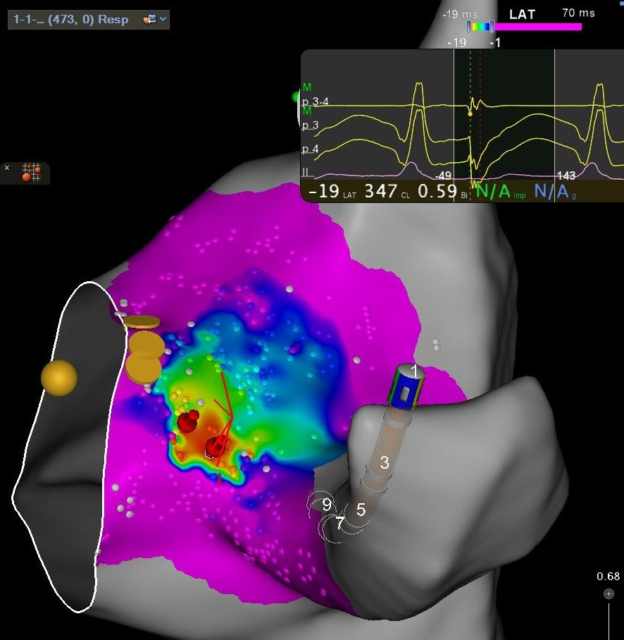
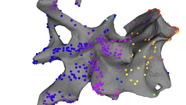
Before these abnormal signal pathways can be ablated, they must be located. This is done with an electrogram: a set of measurements of electrical activity made by placing electrodes directly on the surface of the heart. Standard mapping requires mapping out dozens or even hundreds of points on the cardiac surface. Each point must be identified, verified, and recorded. The recorded data are interpreted by computer to create a local activation time (LAT) map, which shows the flow of signals through the heart tissue.
The mapping process must be repeated after each ablation, further adding to the duration of the operation. This creates a dilemma for the operator: increasing the patient’s surgical risk by taking the time to make a thorough and accurate map versus potentially missing pathways that need ablation.
For this reason, Dr. Mehdi Razavi and his colleagues in the THI Department of Electrophysiology Clinical Research and Innovations collaborated with researchers at Rice University’s Department of Electrical and Computer Engineering to develop a better computer algorithm for creating LAT maps. This algorithm, called MAGIC-LAT, uses a more sophisticated interpolation process than the standard algorithm, which enables MAGIC-LAT to generate a map from fewer points. This shortens the intraoperative time required for mapping. Furthermore, the algorithm generates a more accurate map than standard mapping does.
In addition, when the MAGIC-LAT algorithm creates the map, it uses a different color scheme that is easier to interpret than the color palette used in standard maps.
The researchers published their work through IEEE Xplore after presenting the new algorithm at the 2021 55th Asilomar Conference on Signals, Systems, and Computers in Pacific Grove, CA.
“Because mapping has to be done repeatedly during an ablation procedure, an algorithm that shortens the mapping process can cut down the procedure time considerably,” says Dr. Razavi. “That and the improvement in accuracy will mean less risk and better outcomes for patients.”
Hellar J, Cosentino R, John MM, Post A, Buchan S, Razavi M, Aazhang B. Graph-Based Interpolation of Local Activation Time on the Cardiac Surface. 2021 55th Asilomar Conference on Signals, Systems, and Computers, 2021, pp. 274-278, doi: 10.1109/IEEECONF53345.2021.9723133
News Story By Stephen N. Palmer, PhD, ELS
Featured images from: J. Hellar et al., “Graph-Based Interpolation of Local Activation Time on the Cardiac Surface,” 2021 55th Asilomar Conference on Signals, Systems, and Computers, 2021, pp. 274-278, doi:10.1109/IEEECONF53345.2021.9723133.